A single eye donor can restore or improve the sight of up to five people through different uses of the donated tissue.
Body & Brain
Can a human eye be donated after death?
Can an Eye Be Donated?
Every year, tens of thousands of people have their sight restored by donated tissue from someone who died. But in almost every case, the donated tissue is not what most people picture when they think of donating an eye.
On this page
The short answer
Specific parts of the eye, particularly the cornea and sclera, can be donated and transplanted successfully. The whole eye as a complete organ cannot currently be transplanted because the optic nerve cannot be reconnected.
Eye banks retrieve eyes from donors within hours of death, evaluate the tissue quality, and process specific layers for transplantation. The cornea, the clear front surface of the eye, is the most commonly transplanted part and restores sight in thousands of patients annually. Modern surgical techniques can transplant individual layers of the cornea rather than the full thickness, dramatically improving outcomes. The retina and optic nerve, which connect to the brain, remain outside current transplant capability.
At a glance
Donation begins with careful tissue screening, not whole-eye replacement.
Eye donation leaves the donor's body without eyes, which is disfiguring.
The cornea, a transparent disc of tissue roughly the size of a shirt button, has no blood vessels and is immunologically privileged, meaning the body is far less likely to reject it than almost any other transplanted tissue. This quirk of anatomy has made corneal donation one of the most successful and routine forms of tissue transplantation in medicine.
Imagine being able to repair a fogged lens on a camera without replacing the camera body. Modern eye donation is closer to precision optical component replacement than the wholesale organ transplant most people imagine.
Behind the science
The story behind it
The First Corneal Transplant, 1905
Eduard Zirm, an Austrian ophthalmologist, performed the first successful human corneal transplant in 1905, taking a cornea from an 11-year-old boy whose eye had been removed due to injury and transplanting it into a farm laborer who had been blinded by chemical burns.
The patient recovered useful vision and retained it for years. Zirm's operation, which preceded the discovery of blood groups and the development of anti-rejection drugs by decades, succeeded because the cornea's lack of blood vessels meant the immune system never launched the full rejection response that would have destroyed any other transplanted tissue.
The first successful organ or tissue transplant in human history succeeded partly because the surgeon, perhaps without fully understanding why, chose the one tissue in the body that the immune system was least likely to attack.
Development of DMEK by Melles et al., 2006
Dutch ophthalmologist Gerrit Melles developed the technique of transplanting only the Descemet membrane and endothelial layer of the cornea, a mere 10 to 15 microns of tissue, rather than full-thickness corneal buttons.
Patients achieved better final visual acuity, recovered faster, and had significantly lower rejection rates than with previous techniques. DMEK is now the preferred approach for endothelial diseases like Fuchs' dystrophy in most major centers worldwide.
Diagram
Can an Eye Be Donated: the idea in one diagram
Eye donation works because transplant medicine uses specific eye tissues, especially the cornea, rather than replacing the whole eye. The optic nerve still cannot be reconnected, but corneal layers can be recovered, preserved, matched to the recipient's disease, and transplanted with high success.
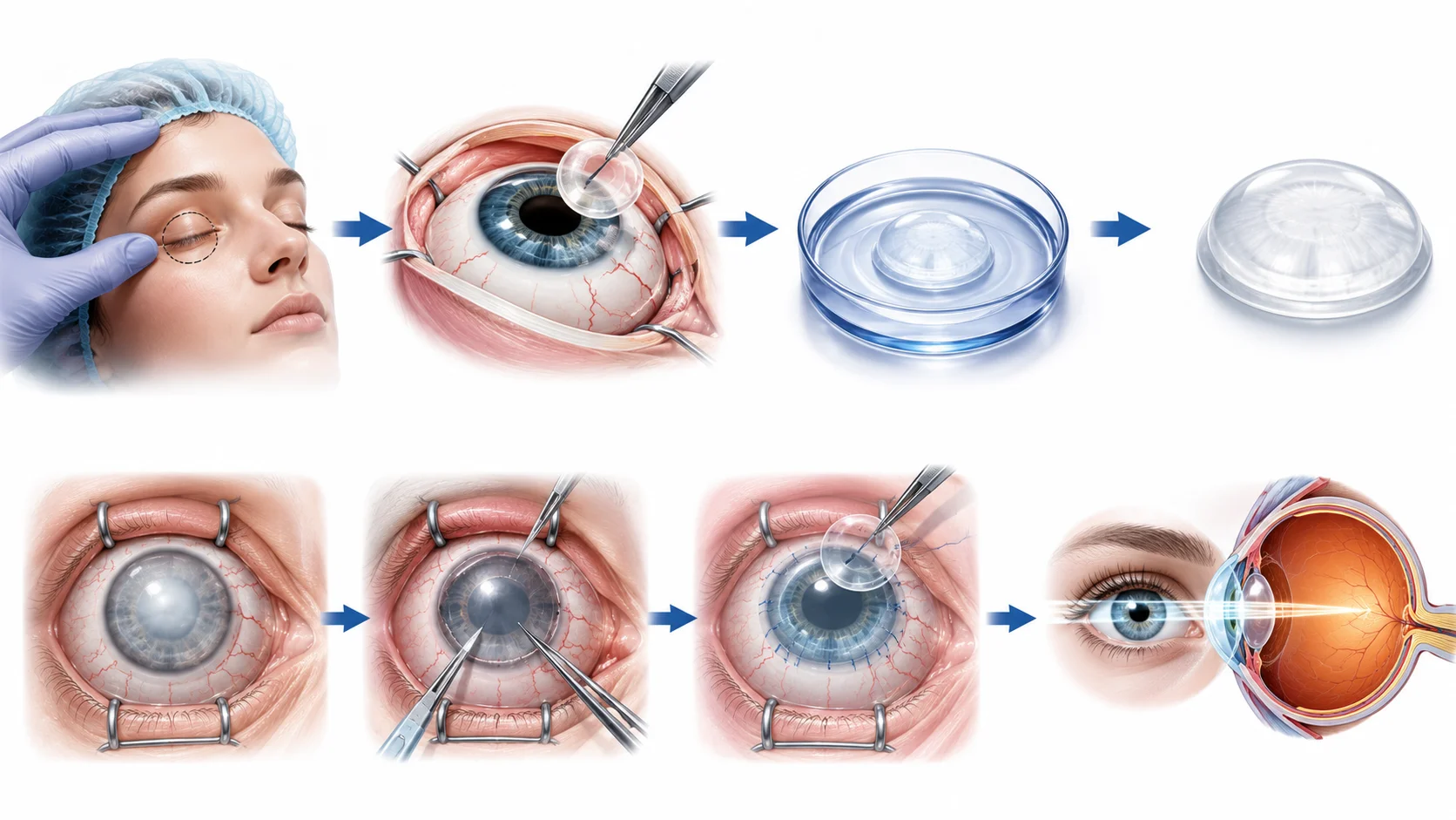
- 1Donor tissue is recovered
Donation begins with careful tissue screening, not whole-eye replacement.
- 2The needed layer is prepared
Most modern corneal donation is targeted layer repair.
- 3The recipient's damaged tissue is replaced
Sight is restored by repairing the eye's clear optical surface.
How it works
The mechanism
Eye donation works because transplant medicine uses specific eye tissues, especially the cornea, rather than replacing the whole eye. The optic nerve still cannot be reconnected, but corneal layers can be recovered, preserved, matched to the recipient's disease, and transplanted with high success.
- Step 01
Donor tissue is recovered
After death, an eye bank retrieves and evaluates the donated eye tissue under sterile conditions, checking corneal clarity, cell density, safety, and suitability for transplant.
It is like inspecting an optical lens before deciding whether it can be used in a precision repair.
Donation begins with careful tissue screening, not whole-eye replacement.
- Step 02
The needed layer is prepared
Depending on the recipient's condition, technicians may prepare a full-thickness corneal graft or only a thin endothelial layer such as DMEK tissue.
Modern corneal surgery can replace one damaged pane in a layered window instead of replacing the whole window.
Most modern corneal donation is targeted layer repair.
- Step 03
The recipient's damaged tissue is replaced
The surgeon removes the diseased corneal tissue and positions the donor tissue so light can again pass cleanly through the front of the eye.
The repair is closer to changing a camera lens element than transplanting an entire camera.
Sight is restored by repairing the eye's clear optical surface.
Surprises
Facts worth sitting with
A single eye donor can restore or improve the sight of up to five people through different uses of the donated tissue.
Surprising facts
What seems backwards
Worth comparing
In everyday life
Easy to get wrong
Pressure points
Where the simple answer gets interesting
Why can't the whole eye be transplanted?
The optic nerve, which connects the retina to the brain, cannot currently be reconnected after severance. The retina processes visual information and transmits it as electrical signals through the optic nerve to the visual cortex. Cutting and reattaching the optic nerve destroys the connection irreversibly. Without optic nerve reconnection, a transplanted eye would be anatomically present but functionally blind.
The same neural reconnection problem that prevents brain transplantation also prevents whole-eye transplantation, just with a smaller but still unresolvable number of nerve fibers.
What is a DMEK transplant and why has it changed corneal surgery?
DMEK (Descemet Membrane Endothelial Keratoplasty) is a technique that transplants only the innermost cellular layer of the cornea, approximately 10 to 15 microns thick, from a donor. It replaces only the diseased endothelial cells rather than the full corneal thickness. The technique produces dramatically faster visual recovery, much lower rejection rates, and better final visual acuity than previous full-thickness transplants.
Modern corneal transplantation has become so precise that surgeons are transplanting tissue layers thinner than a human hair with reliable success.
Can corneal tissue be grown in the lab?
Research into bioengineered corneas is active and producing promising results. Lab-grown corneal endothelial cells have been transplanted successfully in small clinical trials. Collagen-based scaffolds seeded with corneal cells have been tested in humans in some countries. A fully functional synthetic cornea that matches donor tissue in all properties does not yet exist but is an active research goal given the worldwide shortage of donor corneas.
There are 12 million people worldwide waiting for a corneal transplant with no available donor. A bioengineered solution would be one of the most impactful interventions in global ophthalmology.
Clarify
Myth and reality
Myth
Eye donation leaves the donor's body without eyes, which is disfiguring.
Reality
Eye banks remove the cornea or the entire eye under sterile conditions and can restore the donor's appearance with prosthetic shells before the funeral. The process is not visible at a viewing.
Evidence
Standard eye donation protocols specifically address cosmetic concerns to maintain the option of open-casket funerals.
Myth
Doctors might not try as hard to save you if you are a registered eye donor.
Reality
This is the most common misconception that discourages registration. Eye banks are completely separate from emergency and critical care medicine. The decision to declare death and the decision to request donation are handled by different teams under strict protocols.
Evidence
Legal and hospital protocols in all countries with organized donation systems require that the care team and the donation team operate independently.
Looking closer
The deeper pattern
The success of corneal donation rests on a structural biological anomaly: the cornea evolved to be avascular to maintain optical clarity, and that same absence of blood vessels eliminated the immune surveillance that causes rejection in other transplanted tissues.
One way to think about it
Evolution made the cornea transparent by removing its blood supply, and in doing so, accidentally made it the most transplant-friendly tissue in the body. Biology's optical solution became surgery's ethical windfall.
The properties most useful for one purpose sometimes create unexpected advantages in entirely different contexts.
Limits
Where the explanation bends
Keratoconus: corneal thinning in young adults.
Keratoconus is a condition where the central cornea thins and bulges forward into a cone shape, severely distorting vision. It affects roughly 1 in 2,000 people, often developing in the teenage years. In severe cases, corneal transplantation is the only treatment. These younger patients often receive full-thickness or anterior lamellar transplants because they need structural replacement, not just endothelial cell restoration.
Keratoconus makes corneal transplantation necessary in otherwise healthy young people, and the long-term success of their transplants is a testament to the cornea's immunological privilege.
In daily life
What this helps you notice
Registering as an eye donor takes approximately two minutes and could restore the sight of up to five people. The gap between available donor corneas and patient need is driven almost entirely by low registration rates, not by medical or logistical barriers.
Countries with opt-out donation systems, where citizens are presumed to consent unless they actively withdraw, have significantly higher donation rates and shorter waiting lists than opt-in systems.
Remember
Key takeaways
- 1
Parts of the eye, especially the cornea, can be donated and routinely restore sight.
- 2
The whole eye cannot be transplanted because the optic nerve cannot be reconnected.
- 3
The cornea's lack of blood vessels makes it uniquely resistant to immune rejection.
- 4
A single donor can help restore or improve the sight of up to five people.
- 5
Global demand for corneal transplants exceeds supply 70 to 1, primarily due to low donor registration.
The first tissue ever successfully transplanted between humans was chosen almost accidentally for the property that made it transparent: it had no blood supply, no immune surveillance, and thus, no reason to reject a stranger's cells.
The cornea evolved to be clear. To be clear, it gave up its blood vessels. And in giving up those vessels, it became the most generous tissue in the body: replaceable, shareable, capable of being passed from the dead to the living with a precision and a success rate that no other organ has matched. Evolution designed an optical surface. It inadvertently designed a gift.
Read around the idea
More questions with the same curious pull
Nearby doors from the TinyThat archive, chosen by topic, intent, and reader curiosity.
Body & Brain
How the Eye Works
The eye focuses light onto the retina through the cornea and lens.
Body & Brain
Can the Brain Feel Pain?
The brain controls all pain in your body, yet it cannot feel pain itself. Discover why, and what this strange fact re...
Body & Brain
Does Your Brain Eat Itself?
Your brain really does consume parts of itself. Learn about autophagy, synaptic pruning, and why self-consumption is...